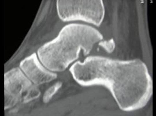
INJURY TO TALUS
The talus can sustain injury in the form of dislocation or fracture. Both these lesions are rare and severe violence is necessary to produce the effect.
DISLOCATIONS OF THE TALUS
The talus is sandwiched between the tibia above and the calcaneum below. Anteriorly the talus forms the talonavicular joint which is a part of the midtarsal joint.
TYPES OF DISLOCATIONS
Usually there are three basic varieties of dislocations.
Subtalar dislocation: The talus loses its attachment from the calcaneum below and the subtalar joint is disorganized. Though the talus maintains its normal relationship within the tibiofibular mortise above, there is disruption of the talocalcaneum and talonavicular articulation below.
Dislocation of the talonavicular joint: The head of the talus is displaced from the concave articular socket of the navicular bone.
Total dislocation of talus: The attachment of talus superiorly with tibia, inferiorly with calcaneum and anteriorly with the navicular bone is completely disrupted. It lies free in an abnormal position.
MECHANISM OF INJURY
All the above-mentioned lesions are produced by the same type of violence. The nature of dislocation depends upon the severity of the force. A fall with the foot in adduction and inversion and the ankle-joint in a plantar-flexed condition can produce a chain of reactions. Firstly the subtalar dislocation is produced by the talus being detached from the calcaneum. When further force is being applied, the talus is finally expelled out of the tibiofibular mortise.
DIAGNOSIS
There is gross deformity and swelling round the ankle-joint. The displaced talus produces a bulging and the skin over it is stretched. In extreme cases the skin covering may get necrosed and the talus may become visible through the wound.
X-ray: X-ray must be carefully interpreted and the nature of dislocation is identified.
TREATMENT
Closed reduction is often successful. Sometimes interposition of tendons may interfere with the process of reduction and operative procedure may be required. At times when the skin over the talus is overstretched, the closed reduction may prove to be a difficult procedure. In this condition it is ideal to resort to operative means, otherwise excessive manipulation may interfere with the viability of the skin. The surgeons use orthopedic screws and other orthopaedic implants for the operative means.
TECHNIQUE
Subtalar dislocation: The basic principles of reduction are to apply force opposite to the direction of adduction and inversion which produced the lesion.
Plantar-flex the ankle: Under general anaesthesia, the patient’s knee is semiflexed and supported in an elevated position. The ankle-joint is forcibly plantar-flexed by the manipulator.
Abduction and eversion of foot: The foot is then abducted and everted. This is opposite to the direction of the force which produced the dislocation.
Total dislocation of talus: The foot is forcibly plantar-flexed and inverted. This produces a space below the tibial articular surface. Pressure is then exerted with both thumbs over the superior articular surface of the talus to replace it into its normal position. During this procedure the ankle-joint should be maintained in a state of plantar flexion.
- Talonavicular dislocation: Closed reduction of the dislocation may be possible. Sometimes even after successful reduction, redislocation may take place.
Closed reduction: Under general anaesthesia the foot is plantar-flexed and everted. Pressure is applied on the head of talus to bring it in apposition with the navicular bone.
Operative procedure: Open reduction and internal fixation by a bone screwpassing through the navicular bone to the head of talus may be necessary. Screws are supplied by orthopedic bone screw manufacturers across the world.
Post-reduction management: A plaster-cast extending from above the knee to the metatarsal head is applied. The leg is kept elevated on a pillow. Check x-ray is taken. The plaster is removed after a period of 8 weeks.


