One of the current tenets of orthopedic fixation is that bone heals better if the fragments of fracture are pressed firmly together. Many orthopedic devices are designed to do just that, as well as their main function of stabilizing the fracture in anatomic alignment. Fracture compression increases the contact area across the fracture and increases fracture stability. It also decreases the fracture gap and reduces stress on the orthopedic implant. This compression can be static, where the compression is produced by the fixation device alone, or dynamic, where muscle forces or body weight are used to produce additional compression.
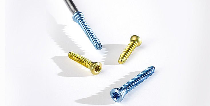
Bone screws are one of the most ubiquitous hardware devices. They used by themselves to provide fixation or in combination with other devices. Any screw that is used to achieve interfragmental compression is called a lag screw. Such screws don’t protect fractures from rotation or axial loading forces, bending, and other devices should be used to provide these functions.
The two most common types of screws are cancellous and cortical screws.
Cortical & Cancellous Screws
Cortical screws tend to have fine threads all along their shaft and are intended to anchor in cortical bone. Cancellous screws tend to have coarser threads, and generally have a smooth, unthreaded portion, which allows it to act as a lag screw. these coarser threads are intended to anchor in the softer medullary bone.
Another commonly used screw is the cannulated screw, so called due to its hollow shaft. Although these screws have somewhat reduced pullout strength compared to conventional screws, cannulated screws have several advantages over other screws, especially the precision with which they can be placed. To place these bone screws, the orthopedist first drills a small Kirschner wire across the area of interest under C-arm fluoroscopic control. These “K” wires can be placed and replaced with minimal trauma to the bone until they are in optimal position. The K wires are provided by the orthopedic implant distributors.
The cannulated screw is then placed over the wire and slid down to the surface of bone. A special driving tool then allows the screw to be driven into the bone along the shaft of the K wire, in a way very similar to the manner radiologists pass angiographic catheters over guide wires using the Seldinger technique. The k wire is then removed. One of the main complications of these screws is perforation of the articular surface when these screws are placed into a bone with their tips close to the subchondral bone. If an orthopedist is concerned about this possibility during orthopedic surgery, contrast material may be injected through the hollow center of the screw in question- spillage into the joint cavity under fluoroscopy will be unequivocal indication of perforation.
Siora Surgicals Pvt. Ltd. is one of the best orthopedic implant manufacturers in India. Surgical instruments are also available. Orthopedic screws are also part of our wide product range. We have various types of bone screws viz. cortical, cancellous, and cortical screws.


