
CT Vs Endoscopy – Quick Overview:
CT scans use X-rays to create images of organs and tissues in the body (e.g., abdominal organs, brain, chest, lungs, heart), and endoscopy reveals only the inner surface of the upper gastrointestinal tract.
CT scans use radiation (X-rays) to generate images, and endoscopy uses a flexible instrument equipped with light and a camera to generate images and biopsies and/or remove polyps from upper gastrointestinal tissue.
Computed tomography is quick, painless, non-invasive, and does not require extensive preparation. Endoscopy, on the other hand, is invasive (the flexible instrument is inserted through the mouth) and generally requires a person to adjust their diet for a short time following the doctor’s instructions.
Conclusion on both:
Both procedures are relatively safe; CT provides radiation (at a safe level). When intravenous contrast media are used to enhance CT images, some people may be allergic or have kidney damage and there is a risk of intestinal perfection and an allergic reaction to anesthesia. In short both are best medical diagnostic tools for human body.
Side effects of colonoscopy
This can include irregular heartbeat, pulmonary aspiration, and/or respiratory depression; if a gastrointestinal perforation occurs, infection and/or bleeding can also occur, and a CT scan can lead to allergic reactions. Dyes and dyes that leach into site IV.
CT scans can be done on people of almost any age, and most endoscopy procedures are done on adults.
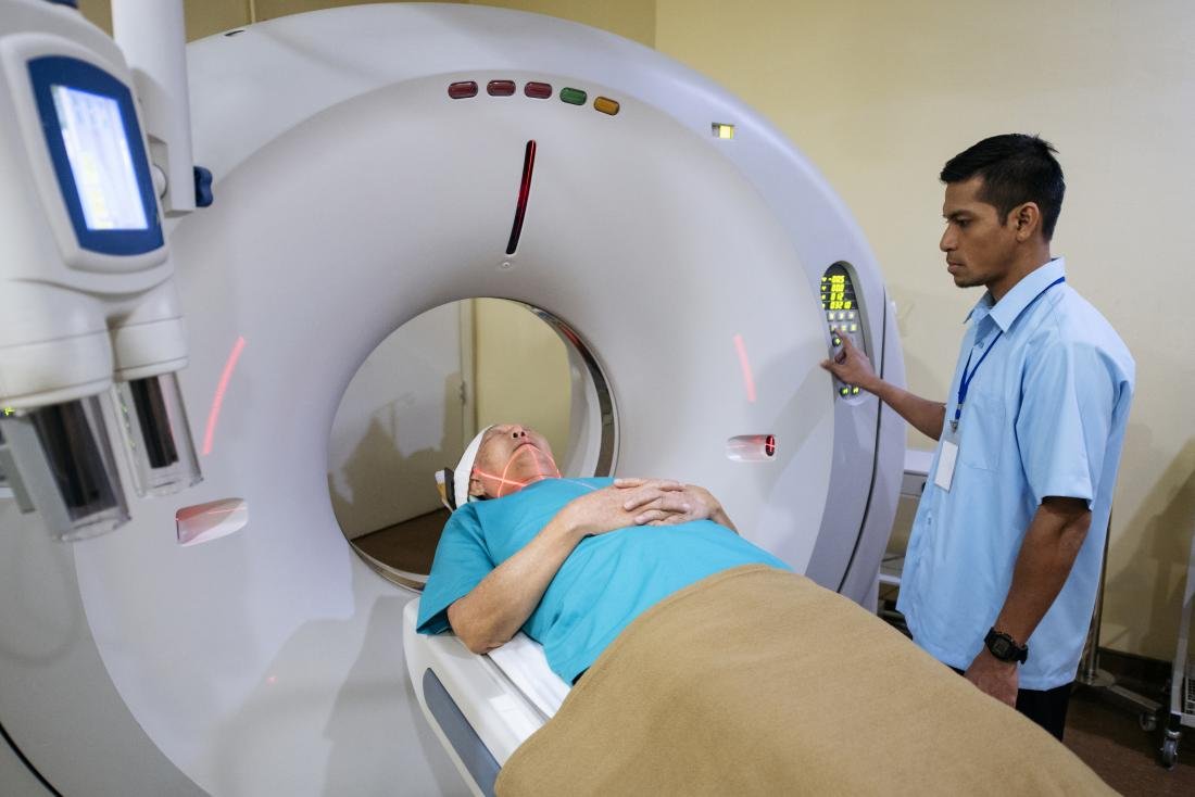
What is a CT scan?
CT scan or CT scan are special x-ray tests that use x-rays and a computer to create cross-sectional images of the body. Computerized axial tomography is called computed tomography.
CT scanners were first installed in 1974. CT scanners have greatly improved patient comfort, as scanning can be performed quickly. The enhancements result in higher resolution images that help clinicians make a diagnosis. For example, CT scans can help doctors see small lumps or tumors that they cannot see on a regular X-ray.
Computed tomography Facts and Figures
- CT images allow the doctor to see the inside of the body like the inside of a loaf of bread would look when cut. This type of X-ray image creates special “images” of parts of the body so that doctors can look directly at the area of interest. CT scans are widely used to examine the brain, neck, spine, chest, abdomen, and pelvis.
- CT is a commonly used technique. Scanners are not only found in hospital X-ray rooms, but also outpatient rooms. When a body get scanned under CT- Scanner, it examines whole body even there no need for any ENT patient to check through any Ophthalmoscope otoscope set, or any cardio patient to go thorough ECG machines.
- CT has revolutionized medicine by allowing physicians to identify diseases that have only been detected in the past, but often during operations or autopsies. CT is non-invasive, safe, and well-tolerated. It provides a very detailed image of many different parts of the body.
- Chest computed tomography is often used to detect abnormalities on a regular chest x-ray. It is also seldom used to consider inflated lymph nodes.
What is an endoscopy procedure?
A procedure called a gastrointestinal endoscopy allows the doctor to view the inside of the digestive tract. This research is done using an endoscope, a flexible fiberglass tube with a small television camera on the end. The camera is connected to a direct view frame or video monitor that displays images on color television. The endoscope allows not only to diagnose gastrointestinal (GI) diseases, but also to cure them.
The gastrointestinal endoscopy procedure can be performed on an outpatient or inpatient basis. A doctor can use the endoscope to evaluate various problems, such as ulcers or muscle spasms. These concerns are not always seen with other imaging tests.
An endoscope has different names depending on what part of the digestive tract the doctor is trying to examine. However, this short article presents the most common use of the term, which is limited to upper gastrointestinal endoscopy, as other procedures are discussed elsewhere (e.g., colonoscopy).
Upper gastrointestinal endoscopy (EGD): This procedure allows to examine the esophagus, stomach and the upper part of the small intestine, the duodenum.
Colonoscopy: With this procedure, the doctor may develop ulcers, inflammation of the skin, membrane intestinal Sli, an abnormal lump and bleeding in the colon or large intestine.
Enteroscopy: Enteroscopy is a diagnostic tool rom it is an improved enteroscope that a doctor can use to view your small intestine.